Wataru Kimura and colleagues at the RIKEN Center for Biosystems Dynamics Research (BDR) in Japan have discovered how the hearts of newborn marsupials retain the ability to regenerate for several weeks. Using this knowledge, the team was able to repair mouse hearts that were damaged a week after birth. The findings, published in the scientific journal Circulation, are expected to contribute to the development of regenerative heart medicines.
Topics:
Topics:
Heart disease is a leading cause of human death and is associated with numerous other secondary illnesses. For humans and other mammals, damaged heart muscle—such as occurs after a heart attack—cannot be naturally repaired because matured heart-muscle cells do not regenerate. As with all tissue regeneration, heart repair requires the birth of new cells, which can only happen through the process of cell division, when one cell becomes two. In most mammalian hearts, muscle-cell division remains possible just after birth, but disappears quickly after a couple days.
However, unlike other mammals, marsupials like kangaroos and koalas are born in an underdeveloped state and many of their internal organs continue to grow after birth, including their hearts. However, not much is known about their capacity for heart regeneration. The team at RIKEN BDR hypothesized that this post-natal heart growth is possible because marsupial heart-muscle cells retain the ability to divide, and that this would allow their hearts to regenerate after injury. They set out to test this theory in the opossum.
They observed that opossum hearts continued to grow for several weeks after birth. They found that the hearts of two-week-old opossums resembled those of one-day-old mice, and that opossum heart-muscle cells continued to divide for weeks after birth. Experimentally induced heart damage at this age repaired itself within a month, indicating that as long as heart cells continue to divide, the heart can be repaired. These results confirmed their hypothesis, and as Kimura notes, “cardiac regeneration for more than two weeks after birth in the opossum is the longest duration observed among mammals investigated to date.”
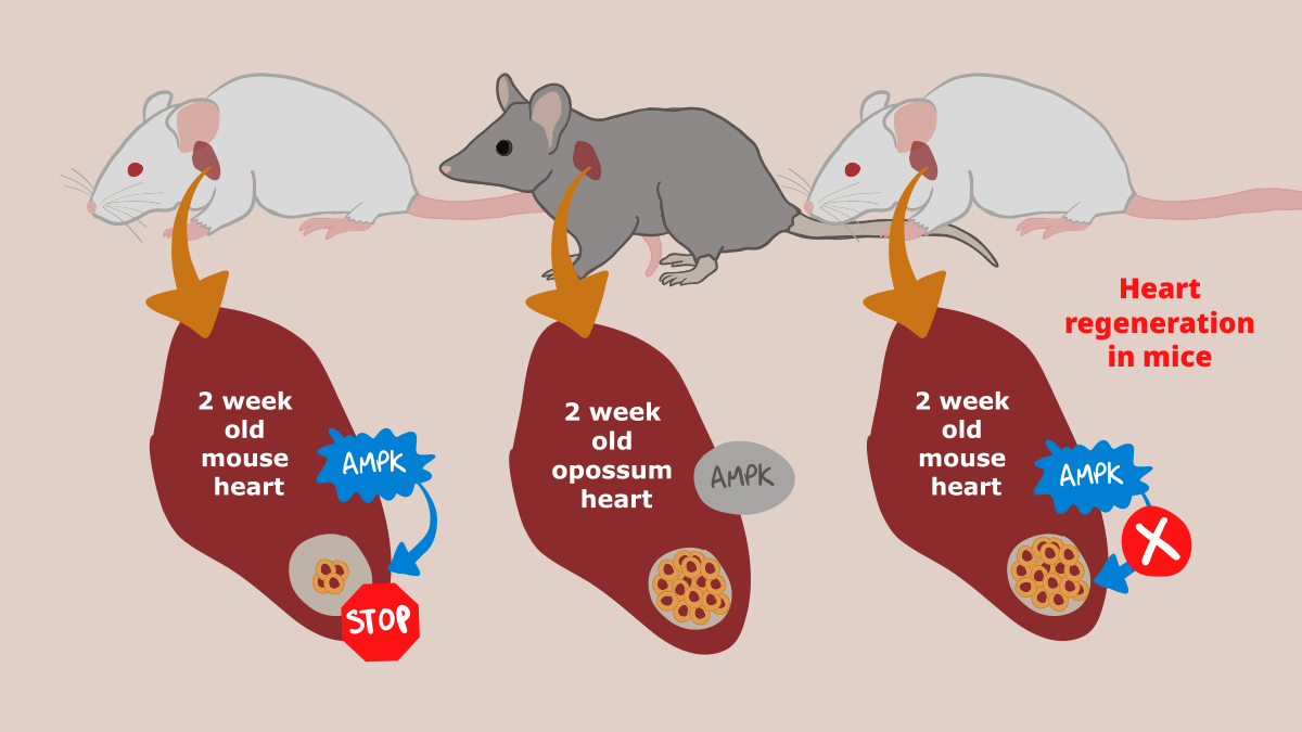
Unlike in mice, in opossums, hearts could still regenerate 2 weeks after birth because AMPK activity was still inactive. Applying this knowledge to mice, researchers were able to prolong the period of time that mouse hearts could regenerate after birth by blocking AMPK activity.
The next step was to figure out how this is possible in opossums but not mice. Gene-expression comparisons showed that two-week-old opossums were similar to mice that were only a few days old. The researchers next looked for changes in gene expression that occurred in both animals around the time that heart regeneration was no longer possible. The common factor was a protein called AMPK. Further experiments showed that activation of AMPK in both mice and opossums coincided with the stoppage of cell division in heart muscle. Therefore, the next hypothesis was that inhibiting AMPK or its ability to work could extend the period during which heart regeneration is possible. As Kimura explains, “if we could exploit the molecular pathway that determines the capacity for cardiac regeneration, we should be able to establish novel therapeutic approaches for treating cardiovascular disease.”
They tested this hypothesis in both opossums and mice, and were successful in both cases. In particular, injecting neonatal mice with AMPK inhibitors allowed hearts that were experimentally damaged a week after birth to regenerate and regain normal function, with minimal scarring. Thus, the researchers were able to use what they learned from marsupials and induce heart regeneration in a regular mammal.
Next on the research agenda is figuring out what triggers AMPK expression at birth in mice but not in opossums. “One important and exciting question is how neonatal marsupials retain regenerative capacity in extrauterine environments, ” says Kimura. “The answers could lead to therapies that can induce heart regeneration in adults.” ??
Heart disease is a leading cause of human death and is associated with numerous other secondary illnesses. For humans and other mammals, damaged heart muscle—such as occurs after a heart attack—cannot be naturally repaired because matured heart-muscle cells do not regenerate. As with all tissue regeneration, heart repair requires the birth of new cells, which can only happen through the process of cell division, when one cell becomes two. In most mammalian hearts, muscle-cell division remains possible just after birth, but disappears quickly after a couple days.
However, unlike other mammals, marsupials like kangaroos and koalas are born in an underdeveloped state and many of their internal organs continue to grow after birth, including their hearts. However, not much is known about their capacity for heart regeneration. The team at RIKEN BDR hypothesized that this post-natal heart growth is possible because marsupial heart-muscle cells retain the ability to divide, and that this would allow their hearts to regenerate after injury. They set out to test this theory in the opossum.
They observed that opossum hearts continued to grow for several weeks after birth. They found that the hearts of two-week-old opossums resembled those of one-day-old mice, and that opossum heart-muscle cells continued to divide for weeks after birth. Experimentally induced heart damage at this age repaired itself within a month, indicating that as long as heart cells continue to divide, the heart can be repaired. These results confirmed their hypothesis, and as Kimura notes, “cardiac regeneration for more than two weeks after birth in the opossum is the longest duration observed among mammals investigated to date.”
Unlike in mice, in opossums, hearts could still regenerate 2 weeks after birth because AMPK activity was still inactive. Applying this knowledge to mice, researchers were able to prolong the period of time that mouse hearts could regenerate after birth by blocking AMPK activity.
The next step was to figure out how this is possible in opossums but not mice. Gene-expression comparisons showed that two-week-old opossums were similar to mice that were only a few days old. The researchers next looked for changes in gene expression that occurred in both animals around the time that heart regeneration was no longer possible. The common factor was a protein called AMPK. Further experiments showed that activation of AMPK in both mice and opossums coincided with the stoppage of cell division in heart muscle. Therefore, the next hypothesis was that inhibiting AMPK or its ability to work could extend the period during which heart regeneration is possible. As Kimura explains, “if we could exploit the molecular pathway that determines the capacity for cardiac regeneration, we should be able to establish novel therapeutic approaches for treating cardiovascular disease.”
They tested this hypothesis in both opossums and mice, and were successful in both cases. In particular, injecting neonatal mice with AMPK inhibitors allowed hearts that were experimentally damaged a week after birth to regenerate and regain normal function, with minimal scarring. Thus, the researchers were able to use what they learned from marsupials and induce heart regeneration in a regular mammal.
Next on the research agenda is figuring out what triggers AMPK expression at birth in mice but not in opossums. “One important and exciting question is how neonatal marsupials retain regenerative capacity in extrauterine environments, ” says Kimura. “The answers could lead to therapies that can induce heart regeneration in adults.” ??
Further reading
Nishiyama et al. (2022) Prolonged Myocardial Regenerative Capacity in Neonatal Opossum. Circulation. doi:10.1161/CIRCULATIONAHA.121.055269
Further reading
Nishiyama et al. (2022) Prolonged Myocardial Regenerative Capacity in Neonatal Opossum. Circulation. doi:10.1161/CIRCULATIONAHA.121.055269

")