The large size of mouse oocytes is shown to create errors when chromosomes divide between daughter cells
Topics:
Topics:
During the first meiotic cell division, in which a parent cell containing two sets of chromosomes produces two daughter cells that each have a single set of chromosomes, immature egg cells known as oocytes make many more mistakes than any other cell type. Such errors can give rise to miscarriages and birth defects. Two RIKEN researchers have now shown experimentally in mice that this propensity to make errors is related to the large size of oocytes1.
“It has long been speculated that the large size of the oocyte is one reason why they are so error prone,” says Tomoya Kitajima from the RIKEN Center for Developmental Biology. “But I wasn’t sure if this could be experimentally tested.” However, when Kitajima saw the excellent micromanipulation skills of co-worker Hirohisa Kyogoku, he was convinced that Kyogoku would be able to test this point.
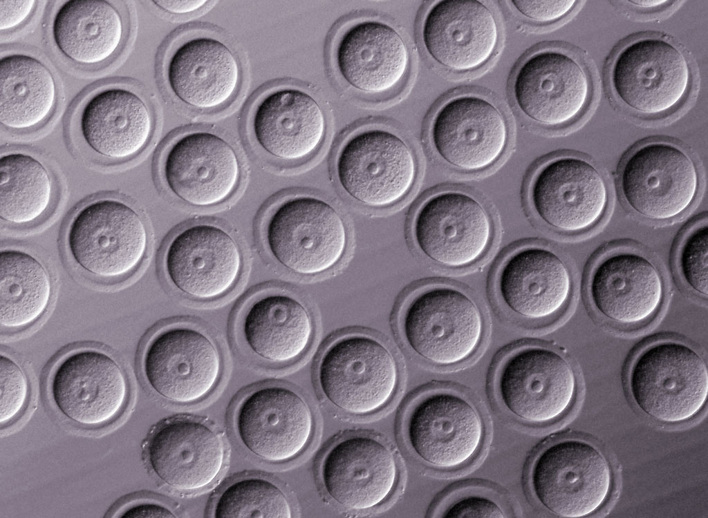
Figure. Scanning electron micrograph of mouse oocytes that clearly shows their nuclei (larger circles inside the cells). © RIKEN CDB
Kyogoku created oocytes that had either half or double the cytoplasm of normal oocytes, by removing half of the cytoplasmic material with a micropipette or fusing two oocytes after removing the nucleus from one of them. The pair then imaged the cells as they underwent cell division to determine whether the volume of cytoplasm affected the accuracy of chromosome segregation.
Cell division is a tightly controlled process. Microtubules emanating from spindle poles attach to condensed chromosomes and align them along a plane at the equator of the spindle (Fig). Proteins that are part of a checkpoint ensure that this alignment is done correctly. If the alignment is not done, division is delayed until correction has taken place.
Kitajima and Kyogoku found that doubled oocytes displayed a variety of defects. In particular, the structure of spindle poles was deformed and thus chromosome alignment at the equator of the spindle was substantially delayed compared to halved and normal oocytes.
In addition to carrying genetic material from the mother, oocytes contain many factors in their cytoplasm that are important for the early development of the embryo after fertilization. The researchers conjecture that factors needed for checking the proper spindle attachment to the chromosomes were diluted by the large cytoplasm. Consequently, the stringency of the checkpoint was compromised and the larger oocytes produced more daughter cells with abnormal distributions of chromosomes.
“We’re now interested in the mechanisms behind this deformation,” says Kitajima. “If we could identify molecules and mechanisms for how the cytoplasm size affects the chromosome segregation machinery, we may find a strategy to make it less error prone.” ?
During the first meiotic cell division, in which a parent cell containing two sets of chromosomes produces two daughter cells that each have a single set of chromosomes, immature egg cells known as oocytes make many more mistakes than any other cell type. Such errors can give rise to miscarriages and birth defects. Two RIKEN researchers have now shown experimentally in mice that this propensity to make errors is related to the large size of oocytes1.
“It has long been speculated that the large size of the oocyte is one reason why they are so error prone,” says Tomoya Kitajima from the RIKEN Center for Developmental Biology. “But I wasn’t sure if this could be experimentally tested.” However, when Kitajima saw the excellent micromanipulation skills of co-worker Hirohisa Kyogoku, he was convinced that Kyogoku would be able to test this point.
Figure. Scanning electron micrograph of mouse oocytes that clearly shows their nuclei (larger circles inside the cells). © RIKEN CDB
Kyogoku created oocytes that had either half or double the cytoplasm of normal oocytes, by removing half of the cytoplasmic material with a micropipette or fusing two oocytes after removing the nucleus from one of them. The pair then imaged the cells as they underwent cell division to determine whether the volume of cytoplasm affected the accuracy of chromosome segregation.
Cell division is a tightly controlled process. Microtubules emanating from spindle poles attach to condensed chromosomes and align them along a plane at the equator of the spindle (Fig). Proteins that are part of a checkpoint ensure that this alignment is done correctly. If the alignment is not done, division is delayed until correction has taken place.
Kitajima and Kyogoku found that doubled oocytes displayed a variety of defects. In particular, the structure of spindle poles was deformed and thus chromosome alignment at the equator of the spindle was substantially delayed compared to halved and normal oocytes.
In addition to carrying genetic material from the mother, oocytes contain many factors in their cytoplasm that are important for the early development of the embryo after fertilization. The researchers conjecture that factors needed for checking the proper spindle attachment to the chromosomes were diluted by the large cytoplasm. Consequently, the stringency of the checkpoint was compromised and the larger oocytes produced more daughter cells with abnormal distributions of chromosomes.
“We’re now interested in the mechanisms behind this deformation,” says Kitajima. “If we could identify molecules and mechanisms for how the cytoplasm size affects the chromosome segregation machinery, we may find a strategy to make it less error prone.” ?
Kyogoku H and Kitajima TS (2017) Large cytoplasm is linked to the error-prone nature of oocytes. Developmental Cell 41, 287–298.e4. doi: 10.1016/j.devcel.2017.04.009
[yuzo_related]
")